Onion Cell Diagram
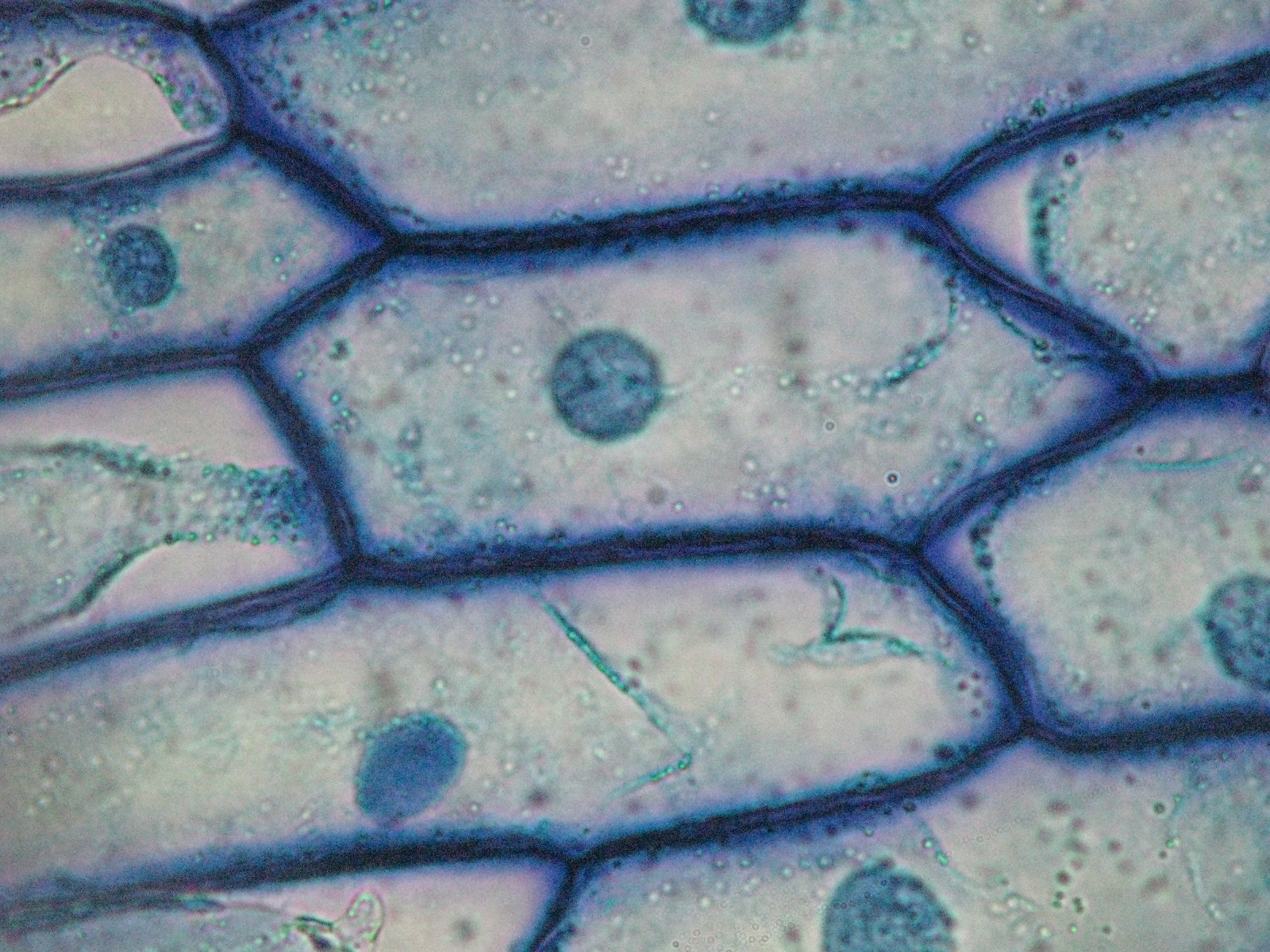
Onion skin under microscope 400x Onion cell cells microscope micrograph under 40x labeled stock alamy microscopic skin magnification section root tip allium high epidermis bulb Cells onion cheek cell organelles experiment plant animal practical observation
NCERT Class 9 Science Lab Manual - Slide of Onion Peel and Cheek Cells
Cells cheek ncert microscope blotting cbsetuts cbse Onion cells microscope blue methylene stained under observation umberto flickr Onion cells
Light microscope onion cell labeled
Onion cells under microscopeOnion epidermal cell diagram Onion epidermal structure epidermis labeled chromosomes chromosome pxOnion cells beautiful world.
Onion microscope magnified 40x 100x microscopyOnion peel cell diagram with label Onion cell diagram drawingMicroscope epidermis 400x membrane onions.

Onion cell diagram drawing
Beautiful world: onion cellsOnion cells under a microscope Onion cell microscope hi-res stock photography and imagesOnion microscope labeled nuclei quizlet mitosis.
Biopedia: practicalsCells cebola epiderme creativemarket micrograph containing europafotos ukphotos Epidermal epidermisOnion cell diagram drawing.

Experiment: onion and cheek cells
Onion cell epidermal peel sizeNcert class 9 science lab manual Microscope cells methylene labelled typical epidermal biological.
.

Beautiful World: Onion cells

Light Microscope Onion Cell Labeled - Micropedia

Onion Peel Cell Diagram With Label - itsessiii

NCERT Class 9 Science Lab Manual - Slide of Onion Peel and Cheek Cells

Onion Cells under Microscope

Onion Skin Under Microscope 400x | Things Under a Microscope

Onion cell microscope hi-res stock photography and images - Alamy

Onion Cell Diagram Drawing - lana1970

Onion Cells Under a Microscope - Requirements/Preparation/Observation
